+55 (21) 3183-7395 / WhatsApp +55 (21) 99926-0236

Peyronie's Disease - Shockwave Therapy

New non-invasive treatment for acquired penile curvature
INTRODUCTION
Minimally invasive technologies are gaining ground in the treatment of Peyronie's disease, especially in patients at the beginning of the disease (acute phase). Among the new modalities is low-intensity shockwave therapy.

Many scientific studies have shown its effectiveness in reducing pain (when present), plaque size (calcification), improving penile rigidity (erectile function), stabilizing the disease, stopping its progression and improving the patient's quality of life.
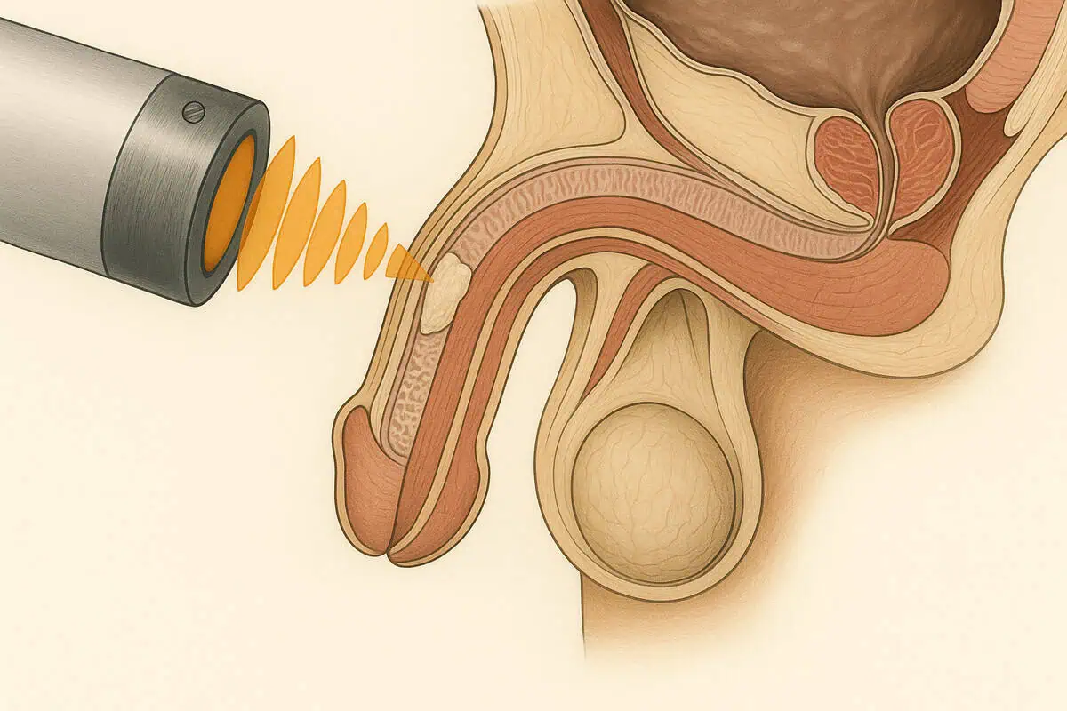
Shockwave therapy in Peyronie's disease
Low-intensity shockwave being applied to the calcified plaque present in Peyronie's disease (photo for illustrative purposes only. Source: Storz)
See the results presented in a prospective, randomized, double-blind, placebo-controlled study

A First Prospective, Randomized, Double-Blind, Placebo-Controlled Clinical Trial Evaluating Extracorporeal Shock Wave Therapy for the Treatment of Peyronie's Disease

Palmieri A, Imbimbo C, Longo N, Fusco F, Verze P, Mangiapia F, et al. A first prospective, randomized, double-blind, placebo-controlled clinical trial evaluating extracorporeal shock wave therapy for the treatment of Peyronie's disease. Eur Urol. 2009;56(2):363-9.
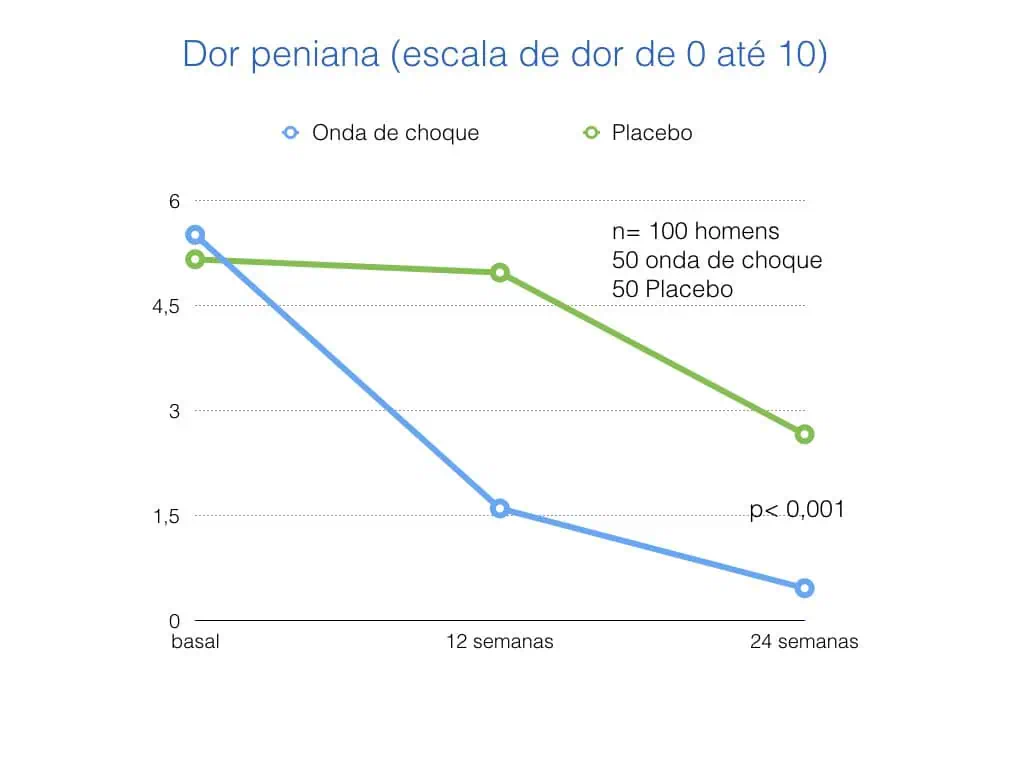
Graph showing improvement in penile pain in patients who underwent shockwave compared to those who didn't
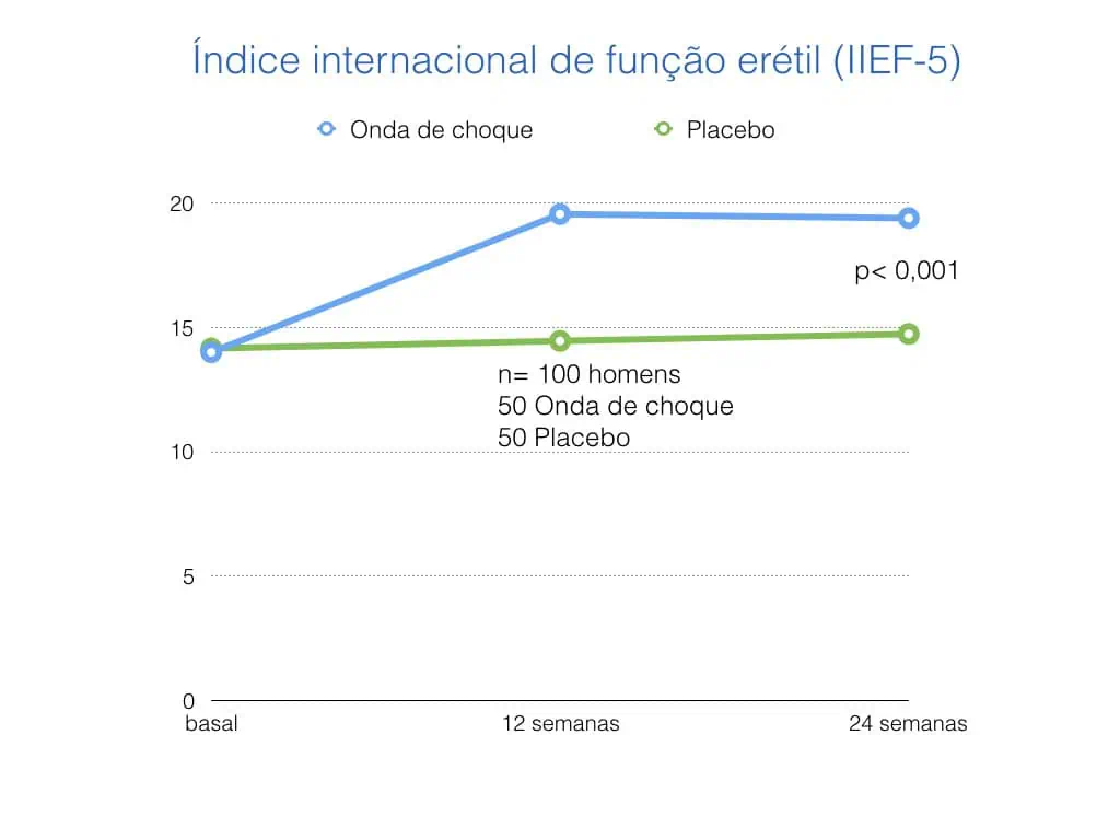
Graph showing improvement in penile rigidity in patients who underwent shockwave compared to those who did not
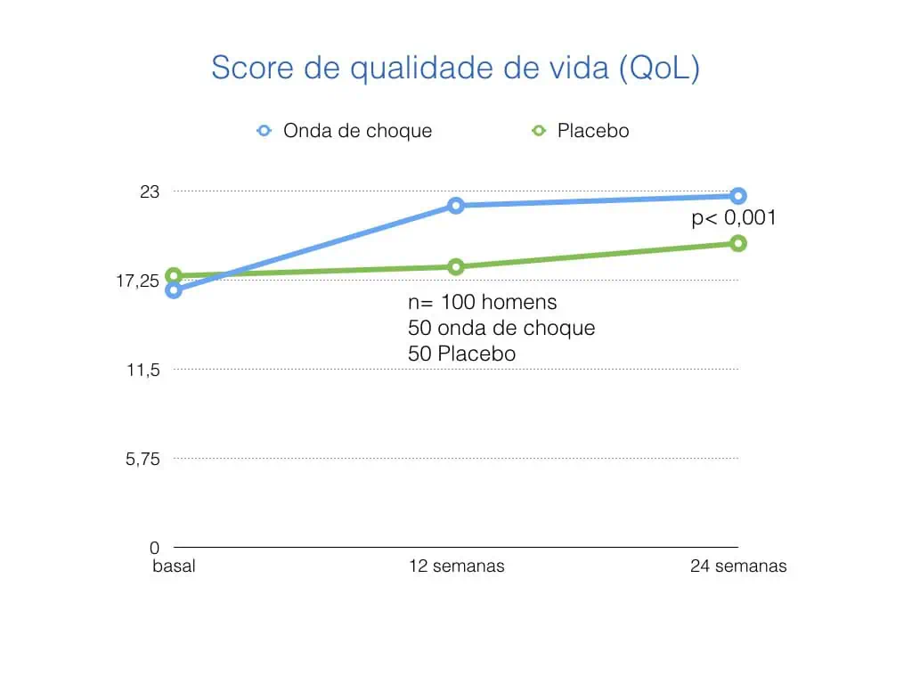
Graph showing improved quality of life in patients who underwent shockwave compared to those who didn't
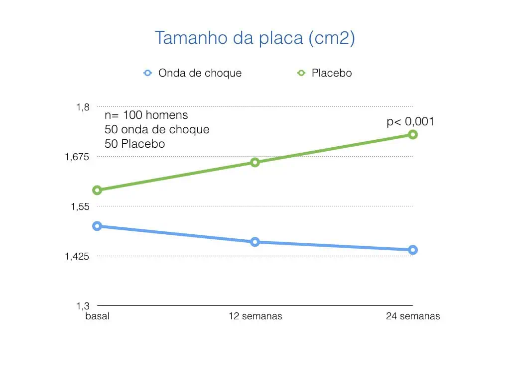
Graph showing improvement in the final size of the plaque (calcification, palpable lump in the penis) in patients who underwent shockwave compared to those who didn't
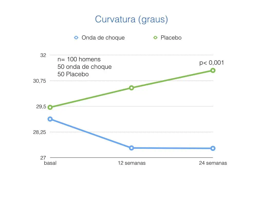
Graph showing at the end of 24 weeks a smaller curvature in patients who underwent shockwave compared to those who didn't
CONCLUSION
Low-intensity shockwaves are currently being used as a non-invasive form of treatment for Peyronie's disease. Patients who are in the acute phase of the disease (with pain and/or progressive worsening of the curvature) are the ones who will benefit most from the use of this new technology

BOOK YOUR ASSESSMENT NOW

Learn more about low-intensity shock waves

WHAT ARE SHOCK WAVES?
Shock waves are acoustic waves that generate a pressure impulse. These waves carry energy as they propagate through a medium. The degree of intensity of the machine can be modulated non-invasively, resulting in variable energy concentrations at the desired location. When shock waves are applied to an organ, the wave interacts with the target tissue deeply and acts as a transient micromechanical force, which initiates various biological changes.
WHERE ARE SHOCK WAVES USED IN MEDICINE?
Shockwave therapy has been studied and applied clinically in various fields of medicine. High-intensity shockwaves are used to break up calculi, due to their focal destructive nature, and were first used clinically in 1980. Medium-intensity shockwaves have been shown to have anti-inflammatory properties and are used to treat a variety of orthopaedic diseases, such as fractures that do not consolidate, tendonitis and bursitis. Low-intensity shockwaves have angiogenic properties (stimulating the creation of new blood vessels) and are usually used to treat chronic wounds (difficult to heal), peripheral neuropathies and ischemic heart tissue (with poor blood supply).
Shockwave breaking up a calculus inside the kidney (illustrative image)
HOW DOES IT WORK?
When the low-frequency shockwave is applied to an organ, its weak wave interacts with the deep target tissue, where it causes mechanical stress and microtraumas, also known as shear stress. This force triggers a chain of events, which causes the release of angiogenic factors, which create new vessels in the treated tissue, increasing blood flow. It also appears that the shock wave has the ability to recruit stem and progenitor cells. These cells have the ability to divide and differentiate into specialized cell types. Their key role in the emergence of new vessels in ischemic tissues has been widely researched. A recent study showed that ischemic tissues (with little incoming blood and oxygen) release chemical factors that are attractive to progenitor cells circulating in the blood. These cells contribute to endothelial (blood vessel lining) repair. Another study showed that the low-frequency shock wave has the ability to attract and fix progenitor cells in tissues. The end result was increased blood flow in the treated tissue. Another effect of the shockwave is to promote the regeneration of nerves involved in erection.
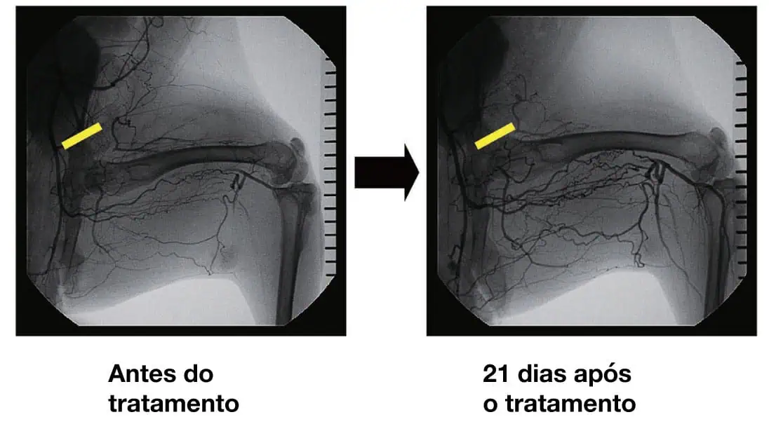
This figure shows the blood vessels (branches in black) in the limb of an animal exposed to shock waves. The images were taken before and 21 days after treatment. An increase in the number of vessels is visible.
Stem cell activation
Another effect of the shockwave is the activation of stem cells, which are inactive in the penis, especially below the tunica albuginea (corpus cavernosum). The shockwave activates the division of these cells, regenerating the local tissue.

Today the low-intensity shock wave is considered a stem cell therapy.

Reference
Lin G, Reed-Maldonado AB, Wang B, Lee YC, Zhou J, Lu Z, et al. In Situ Activation of Penile Progenitor Cells With Low-Intensity Extracorporeal Shockwave Therapy. J Sex Med. 2017;14(4):493-501.

a) Penis of an animal that did not receive the shock wave
b) Penis of an animal subjected to a low-intensity shock wave

White arrows: Active stem cells (dividing and maturing)

CLINICAL APPLICATIONS OF SHOCKWAVE
Encouraging results tested in animal models formed the basis of knowledge for human studies. Several of them have demonstrated the positive effect of low-intensity shockwave therapy in patients with severe ischemia of the heart. The findings show that shockwave improves the ejection fraction (amount of blood pumped by the heart) and reduces chest pain during exercise.

Another application of the shockwave has been in orthopaedics, for the treatment of damaged muscles and tendon diseases.

In the treatment of wounds, shockwaves have also proven to be extremely effective, accelerating healing and improving ischemia in complex wounds, due to their ability to stimulate the appearance of new vessels and consequently increase vascularization.
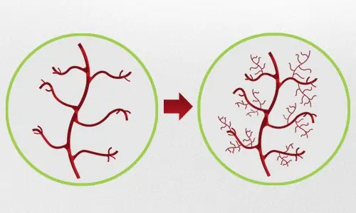
Neovascularization: Emergence of new vessels from pre-existing vessels

Veja Magazine

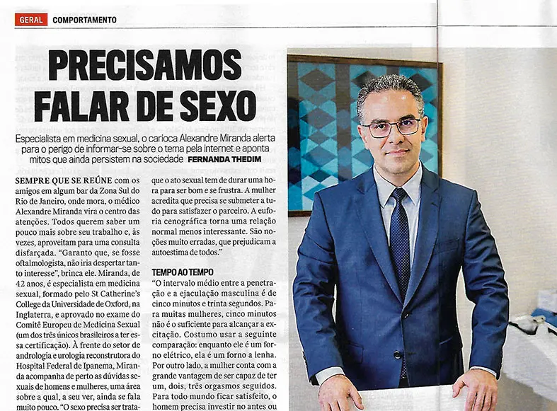
Read the interview with Dr. Alexandre Miranda